Atlas Imagistica's teleradiology service relies on a four-layer technological infrastructure—from image acquisition at partner clinics, through secure transfer and AI-assisted intelligent allocation, to subspecialized reporting—designed to minimize technical barriers so radiologists can focus on accurate diagnosis.
The Technology Behind Atlas Imagistica Teleradiology: A 4-Layer Infrastructure Supporting Every Report
How the complete technological chain of a teleradiology service works, from patient scanning to the delivery of a subspecialized report.
Teleradiology is most often perceived as a radiologist interpreting images remotely. This definition is not incorrect, but it overlooks a critical aspect: the technological infrastructure behind every interpreted study.
A teleradiology service is only as good as the systems it relies on. The quality of a report depends not only on the radiologist’s expertise, but also on how the images reach them, the post-processing tools available, the speed of delivery, and the clinical context accompanying the study.
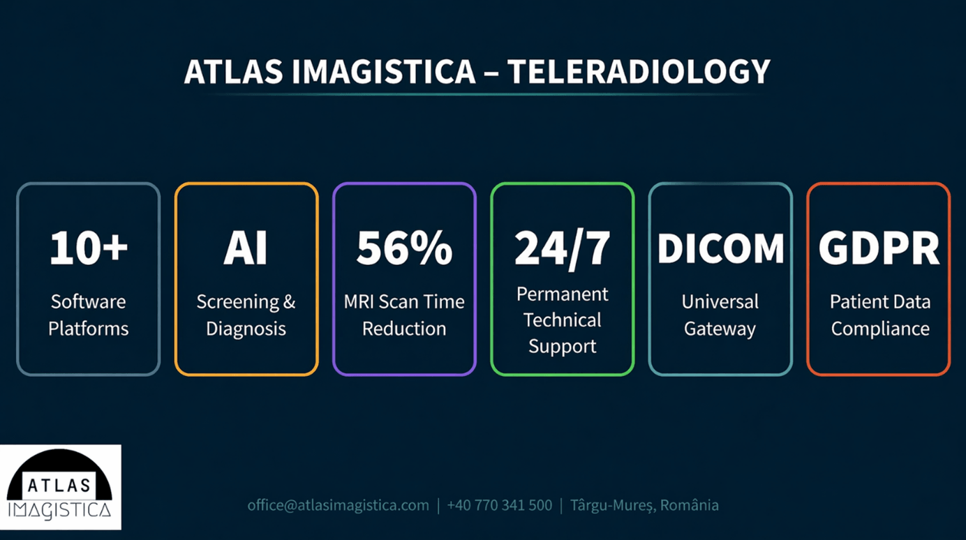
At Atlas Imagistica, the technological architecture is built on four distinct layers, each with a precise role in the chain from image acquisition to the delivery of a subspecialized report. The guiding philosophy is straightforward: the fewer technological barriers a radiologist faces, the more cognitive energy can be invested in diagnostic interpretation. The quality of the report directly reflects this.
Level 1: Image Acquisition at the Partner Clinic
The starting point is always the partner clinic. The patient is scanned using the clinic’s own equipment, whether it is a Siemens CT, a GE MRI, or imaging equipment from another manufacturer. Atlas Imagistica works with any system through the universal DICOM standard, eliminating compatibility issues.
Connection to the platform is achieved via a DICOM gateway (hardware or software), installed at the partner clinic with minimal cost. Once configured, transmitting an imaging study is just one click away, directly from the clinic’s DICOM viewer. It does not require changes to the existing workflow and does not involve complex technical configurations for the clinic’s team.
Level 2: Secure Transfer via TeleradiologyNET
After acquisition, the images are automatically transmitted through TeleradiologyNET, Atlas Imagistica’s proprietary teleradiology platform, via an SSL-encrypted connection or a dedicated VPN channel. DICOM and HL7 standards ensure full interoperability with the partner clinic’s RIS/PACS systems.
A key differentiator: relevant clinical information (medical history, referral diagnosis, medication) is integrated into the platform. The radiologist receives not only images, but also the clinical context necessary for an informed interpretation. The transfer typically takes approximately 10 minutes from the completion of the scan.
Level 3: Intelligent Allocation, AI, and Post-Processing
Once in the platform, the study is handled by the automatic allocation system. The algorithm identifies the most suitable radiologist based on three criteria: availability (who is on shift), subspecialization (cardiology, neuroradiology, MSK, ENT, etc.), and patient continuity (if the same radiologist has interpreted previous studies for the same patient). Urgent cases are flagged with priority and appear at the top of the worklist.
Post-processing: enterprise-level tools
Radiologists have access to a suite of post-processing tools that cover virtually all imaging subspecialties. Siemens Syngo.via generates the necessary reconstructions (cardiac, vascular, neuroimaging) within seconds, while Syngo.Carbon, in its zero-footprint version (browser-based), enables fast reporting without reliance on locally installed software. GE AW Server expands cardiac and vascular analysis capabilities, and PixelData viewers (2D, 3D, and Web) provide flexible visualization adapted to each study type.
In addition to these core platforms, Atlas Imagistica uses specialized systems: Quantib Brain for quantitative brain analysis, PROView for prostate evaluation, Circle Cardiovascular for cardiac imaging, OncoQuant for oncology, and TAVI Analysis for valve intervention planning. A technology that reduces MRI scan time by up to 56% is also available.
Artificial Intelligence: A Support Tool, Not a Replacement
The AI systems integrated at Atlas Imagistica function as a screening filter, not as a substitute for clinical judgment. Within dedicated workflows, AI is active in chest radiography (detecting conditions such as pneumonia, pneumothorax, pulmonary nodules, cardiomegaly, pleural effusions, or rib fractures) and chest CT (pulmonary nodules, emphysema, mediastinal pathology).
An advanced AI imaging screening system covers five clinical areas:
• Spine/chest/abdominal MRI: detection of osteoporosis, bone lesions, abdominal volumetry, liver fat quantification
• CT: identification of pulmonary nodules and coronary calcifications (Agatston score)
• Knee MRI: evaluation of synovial fluid, cartilage, and ICRS score
• Brain MRI: detection of aneurysms, brain volumetry with 34 quantitative markers, Fazekas score, and white matter lesions
• Mammography: breast lesions and arterial calcifications as cardiovascular risk markers
Results are automatically generated and integrated into the reporting workflow, without additional manual steps. The concrete impact: reduced rate of missed lesions and shorter reporting times. Every AI result is reviewed and validated by a subspecialized radiologist.
Level 4: Report Delivery and Partner Support
The finalized report is delivered to the partner clinic’s teleradiology portal and uploaded directly and securely into its PACS system. The clinic accesses the report together with the original images, without additional steps. If the referring physician requires clarification or wishes to discuss a case, communication with the radiologist is facilitated via the helpdesk team, available by phone, chat, or audio/video.
In addition to individual reports, partner clinics have access to customized management reports (scan volumes, monthly statistics) and a complete audit trail with precise timestamps for each interpreted study. Delivery operates 24/7, 365 days a year.
Ongoing Commitment to Innovation
The technological infrastructure described above is not a finished project. Atlas Imagistica continuously runs multiple development initiatives focused on new AI solutions, visualization platforms, and tools designed to streamline radiologists’ workflows. The IT Development team works on in-house products meant to complement external solutions and address the specific needs of the Romanian medical market.
The selection criteria for any new technology remain the same, regardless of vendor or complexity: does it deliver measurable clinical value? Does it reduce the radiologist’s mental workload? Does it improve the partner clinic’s experience? If the answer is yes, the investment is made. This approach explains why the Atlas Imagistica architecture evolves continuously, without compromising the stability expected by partner clinics from a teleradiology service.